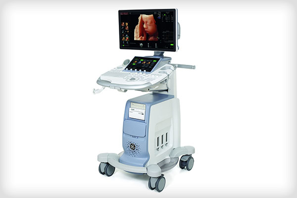
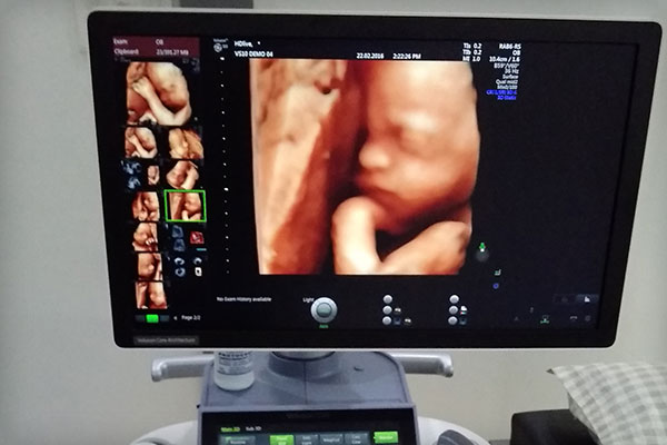
Ultrasound is a safe and painless procedure that is used to produce images of the inside of the body using sound waves. Ultrasound imaging, also called ultrasound scanning or sonography, involves the use of a small transducer (probe) and ultrasound gel placed directly on the skin. High-frequency sound waves are transmitted from the probe through the gel into the body. The transducer collects the sounds that bounce back and a computer then uses those sound waves to create an image. Ultrasound examinations do not use ionizing radiation (as used in x-rays), thus there is no radiation exposure to the patient. Because ultrasound images are captured in real-time, they can show the structure and movement of the body’s internal organs, as well as blood lowing through blood vessels.
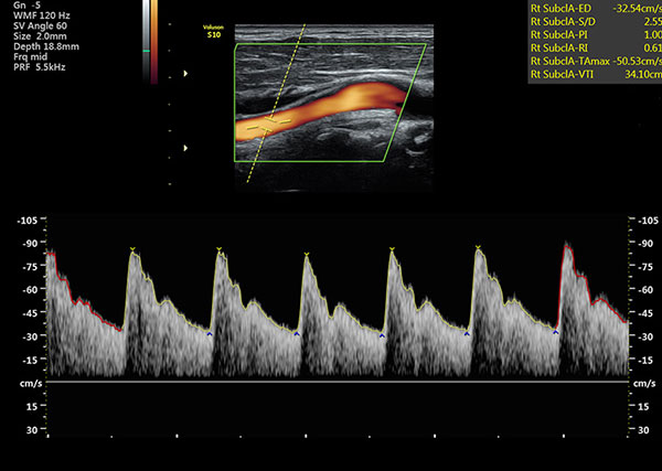
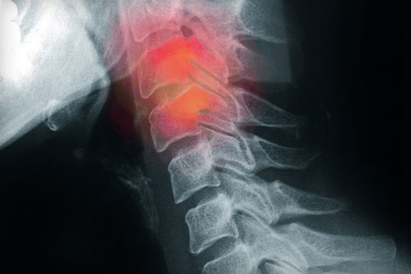
Doppler vascular ultrasound or a color Doppler ultrasound is a special ultrasound technique that allows the physician to see and evaluate blood low through arteries and veins in the abdomen, arms, legs, neck and/or brain (in infants and children) or within various body organs such as the liver or kidneys. Vascular ultrasound produces precise images and measurements of many blood vessels in the body. It can detect diseased vessels and identify a wide variety of changing conditions, enabling the radiologist to make a quick and accurate diagnosis.
The preparations for an ultrasound vary depending on the body part you are having scanned.
For studies involving the liver, gallbladder (the gallbladder contracts when food is digesting, so it is very important to follow these directions so the entire gallbladder can be visualized), spleen and pancreas, eat an early low-fat dinner on the night before your exam and have nothing to eat or drink for 8 to 12 hours prior to your exam.
For studies of your Pelvis you will need to have a full bladder for this exam. Please drink 4-6 glasses of water. Finish drinking water one hour prior to your exam. Do Not Empty your bladder before the exam.
For studies of your Kidneys, you may be asked to drink four to six glasses of water one hour prior to your exam so that your bladder can be visualized.
For studies involving your aorta, please avoid eating for 8 to 12 hours prior to your exam.
There is no preparation if you are having an ultrasound of your Breast, Extremity or other body parts (i.e., Thyroid, Scrotum).
You should wear comfortable, loose-itting clothing for your ultrasound exam. You may need to remove all clothing and jewelry in the area to be examined. Depending on may be asked to wear a gown during the procedure.

You will be free to leave the facility and resume normal activities as your health permits. You will receive your ultrasound report within 30 mins.
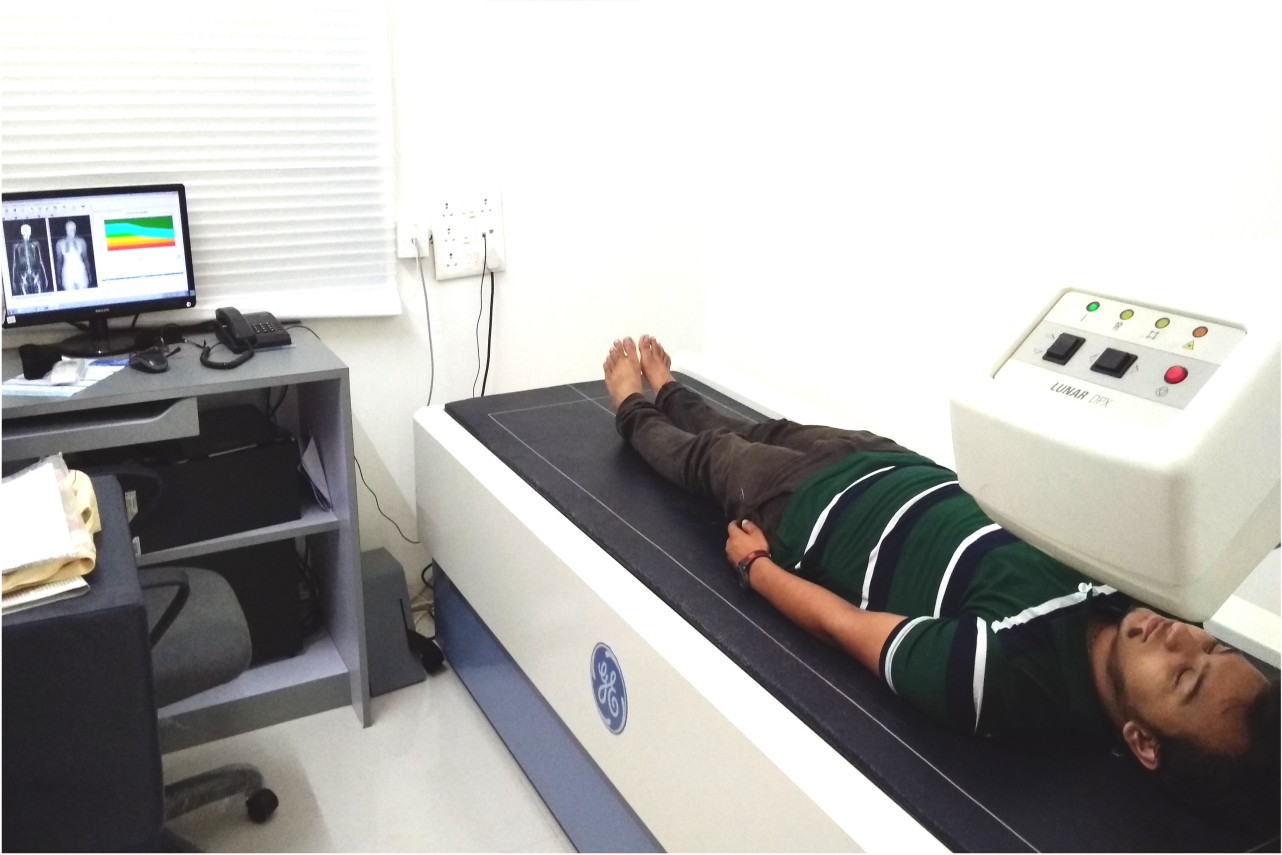
For most ultrasound exams, you are normally asked to lie down on a comfortable exam table where the area to be examined is exposed while the rest of the body is covered.
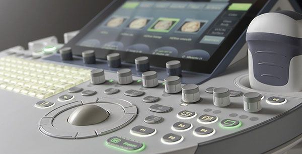
After you are positioned on the table, the radiologist will apply a warm water-based gel to the area of the body being studied. The sonographer will use a small hand held devise called a transducer, which is approximately the size of a bar of soap to complete the exam. Then the sonographer places the transducer on the body and gently moves it back and forth over the area of interest until the desired images are captured.
There is usually no discomfort during the exam when the transducer is pressed against the area being examined.
Once the imaging is complete, the water-based gel will be wiped off your skin, and any portions that are not wiped off will dry to a powder. The ultrasound gel does not stain or discolor clothing